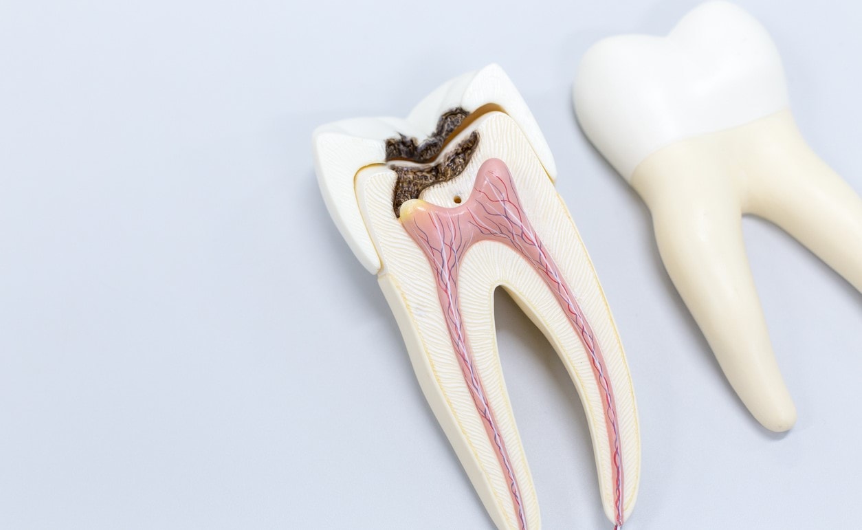
Endodontics consists of treating the contents of the roots, that is, the dental pulp and peri-apical infections (located inside the bone at the tip of the roots).
Treatment is usually performed when the tooth nerve is infected because of a too deep cavity, a large crack in the tooth, or injury.
This is called a root canal.
Why perform this treatment under a microscope?
The endodontist uses diagnostic tools such as a CT scan or 3D imaging, as well a therapeutic tools: a laser and operating microscope.
Because root canals are sometimes not visible to the naked eye, the endodontist uses the microscope to diagnose the presence of inflammation, small cracks, or an infection of the root.
The Operating Microscope
This is an articulated devise on an arm, equipped with great optical features: it has powerful magnification and lighting which eliminates dark areas in the treatment zone.
It also makes it possible to manage the areas that are the most difficult to reach with the naked eye, and also enables precise surgery and a better diagnosis.
The operating microscope makes it possible to detect the root canals, isthmuses, perforation, cracks, lateral canals, and microfractures.
The microscope also makes it possible to retreat certain teeth which could have been condemned.
It is a meticulous treatment: appointments last 1.5 hours and 1 to 3 sessions may be needed to save the tooth.
For more informations or to book an appointment with
Dr Sebastian FERCHERO- Dentist in Nice (France), please contact us by email or call 0033 492 145 145

